 Research Interests:
Research
InterestsMy laboratory develops statistical, image analysis and visualization methods for unraveling basic mechanisms of biology. We are particularly interested in multicellular systems and assembling quantitative system-wide information derived at cellular and subcellular resolution. Read more ..... Curriculum Vitae |
Knowles Lab BioImaging Research LBNL
David William Knowles Cellular and Tissue Imaging Department, Molecular Biophysics & Integrated Bioimaging Division, Lawrence
Berkeley National
Laboratory |
In
the News: 24Jun05: Era of Hope 8Mar06: New Imaging Method ID's Cancer Cells Early more.. 21April06: Early Cancer Detection 2007: Gene Expression stories by BioScienceMag.org, Nature Review Genetics, American Scientist and LBNL 2007: GSA, Drosophila Image Award May 2008: Developmental Cell June 2008: Nature Methods |
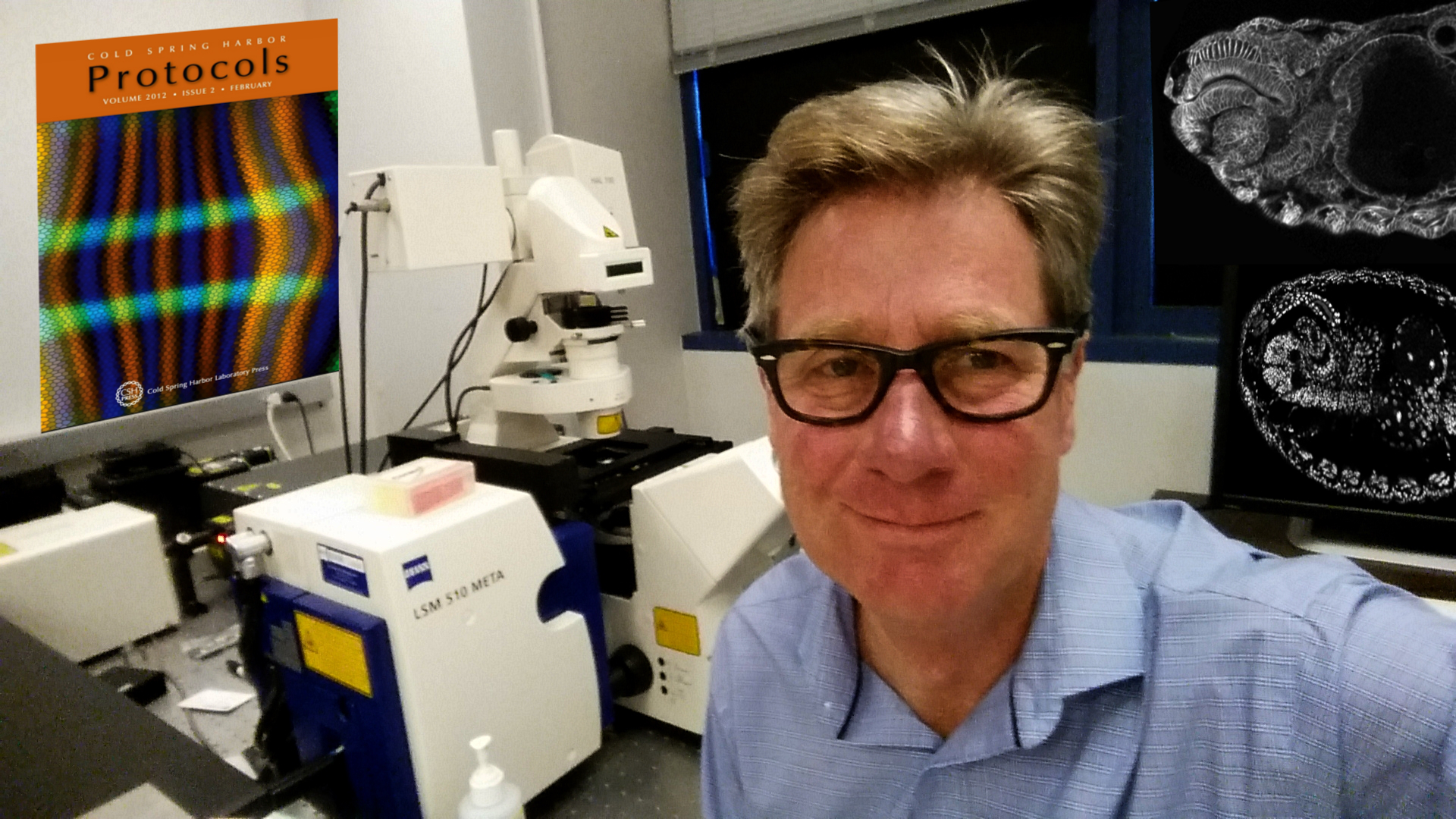


Image-based Screening of Mammary Tumors
The
BioImaging Group is developing methods for turning high
resolution fluorescence images
of human mammary epithelial tissue into tissue-maps which report the
probable nonneoplastic,
premalignant and malignant phenotypes at cellular resolution.

Morphology and Gene Expression in Embryonic Drosophila
The BioImaging Group has created the first computational, quantitative, cellular resolution atlas
of morphology and gene expression for one of the most studied model animals:
Drosophila
melanogaster.

Automated Scientific Analysis Processing (ASAP):
- Fluorescence Image Detection, Segmentation and Quantitation
- Real Time Bright Field Morphology Analysis
- Remote Software Developement
- Machine Independent Implementation
Publications:
Building quantitative, three‐dimensional atlases of gene expression and morphology at cellular resolution
Wiley Interdisciplinary Reviews: Developmental Biology 2013 DOI: 10.1002/wdev.107 Reprint
J Barron, M Biggin, P Arbelaez, D Knowles, S Keranen, J Malik 2013
Volumetric semantic segmentation using pyramid context features
Proceedings of the IEEE International Conference on Computer Vision, 3448-3455 Reprint Supplement
DW Knowles 2012
3D Image-Based Whole Embryo Morphology and Gene Expression Mapping for the Drosophila blastoderm
Cold Spring Harbor Protocols 2012 (2), 150 Reprint
 |
Protocols cover art |
Charless C Fowlkes, Kelly B Eckenrode, Meghan D Bragdon, Miriah Meyer, Zeba Wunderlich, Lisa Simirenko, Cris L Luengo Hendriks, Soile VE Keränen, Clara Henriquez, David W Knowles, Mark D Biggin, Michael B Eisen, Angela H DePace 2011
A conserved developmental patterning network produces quantitatively different output in multiple species of Drosophila
PLoS Genet 7 (10), e1002346 Reprint
Anil Aswani, Soile V.E. Keranen, James Brown, Charless C. Fowlkes, David W. Knowles, Mark D. Biggin, Peter Bickel and Claire J. Tomlin 2010. Nonparametric identification of regulatory interactions from spatial and temporal gene expression data
BMC Bioinformatics 2010, 11:413 doi:10.1186/1471-2105-11-413
Oliver Rubel, Gunther H. Weber, Member, Min-Yu Huang, E. Wes Bethel, Mark D. Biggin, Charless C. Fowlkes, Cris L. Luengo Hendriks, Soile V. E. Keranen, Michael B. Eisen, David W. Knowles, Jitendra Malik, Hans Hagen, and Bernd Hamann 2010
Integrating Data Clustering and Visualization for the Analysis of 3D Gene Expression Data
IEEE Transactions on Computational Biology and Bioinformatics. 2010, Vol 7, No. 1, p64-79 [Reprint]
Stewart MacArthur, Xiao-Yong Li, Jingyi Li, James B. Brown, Hou Cheng Chu, Lucy Zeng, Brandi P. Grondona, Aaron Hechmer, Lisa Simirenko, Soile V.E. Keränen, David W. Knowles, Mark Stapleton, Peter Bickel, Mark D. Biggin and Michael B. Eisen 2009. Developmental roles of 21 Drosophila transcription factors are determined by quantitative differences in binding to an overlapping set of thousands of genomic regions
Genome Biology 2009, 10:R80
Gunther H. Weber, Oliver Rubel, Min-Yu Huang, Angela H. DePace, Charless C. Fowlkes, Soile V. E. Keranen, Cris L. Luengo Hendriks, Hans Hagen, David W. Knowles, Jitendra Malik, Mark D. Biggin,and Bernd Hamann 2009
Visual Exploration of Three-dimensional Gene Expression Using Physical Views and Linked Abstract Views
Computational Biology and Bioinformatics, IEEE/ACM, Transactions on. 6:296-309, 2009 [Reprint]
Charless C. Fowlkes, Cris L. Luengo Hendriks, Soile V.E. Keranen, Gunther H. Weber,1,5 Oliver Rubel,Min-Yu Huang, Sohail Chatoor, Angela H. DePace, Lisa Simirenko, Clara Henriquez, Amy Beaton, Richard Weiszmann, Susan Celniker, Bernd Hamann, David W. Knowles, Mark D. Biggin, Michael B. Eisen, and Jitendra Malik 2008
A quantitative spatiotemporal atlas of gene expression in the Drosophila blastoderm
Cell 133:364-374 April 18, 2008 [Reprint]
 |
A registration technique is described that takes image-based data from hundreds of Drosophila blastoderm embryos and builds a model spatiotemporal embryo atlas from which we recover known gene-regulatory interactions and predict hundreds of new ones. |
Xiaoyong Li, Stewart MacArthur, Richard Bourgon, David Nix, Daniel A. Pollard, Venky N. Iyer, Aaron Hechmer, Lisa Simirenko, Mark Stapleton, Cris L. Luengo Hendriks, Hou Cheng Chu, Nobuo Ogawa, William Inwood, Victor Sementchenko, Amy Beaton, Richard Weiszmann, Susan E. Celniker, David W. Knowles, Tom Gingeras, Terence P. Speed, Michael B. Eisen and Mark D. Biggin 2008
Transcription Factors Bind Thousands of Active and Inactive Regions in the Drosophila Blastoderm
PLoS Biol. 6, e27 (2008)
Cris L. Luengo Hendriks, Soile V. E. Keränen, Mark D. Biggin and David W. Knowles 2007
Automatic channel unmixing for high-throughput quantitative analysis of fluorescence images
Optics Express 15(19):12306-12317
Fuhui Long, Hanchuan Peng, Damir Sudar, Sophie A. Lelièvre, and David W. Knowles 2007
Phenotype Clustering of Breast Epithelial Cells in Confocal Images based on Nuclear Protein Distribution Analysis
BMC Cell Biology 2007, 8(Suppl 1):S3
Gurushankar Chandramouly, Patricia C. Abad, David W. Knowles, and Sophie A. Lelièvre 2007
Nuclear organization and tissue polarity cooperate to control cell fate in mammary acini
J. Cell Sci. 120, 1596-1606 (2007) [Reprint]
Luengo Hendriks C.L. & Knowles D.W. 2007
Comments on the paper ‘A novel 3D wavelet-based filter for visualizing features in noisy biological data’, by Moss et al.
J. Microscopy, 225 (1): 104–107 [Reprint] --> [See Reply by Moss et al]
Patricia C. Abad,
NuMA Influences Higher Order Chromatin Organization in Human Mammary Epithelium
Mol. Biol. Cell, 18:348-361 [Reprint]
Soile VE Keranen , Charless C Fowlkes , Cris L Luengo Hendriks , Damir Sudar , David W Knowles , Jitendra Malik and Mark D Biggin 2006
3D morphology and gene expression in the Drosophila blastoderm at cellular resolution II: dynamics
Genome Biology 2006, 7:R124 Reprint
 |
A new spatio-temporal coordinate framework for studying three-dimensional patterns of gene expression in the Drosophila blastoderm is presented that takes account of previously undetected morphological movements. |
 |
A suite of methods that provide the first quantitative three-dimensional description of gene expression and morphology with cellular resolution in whole Drosophila embryos is described. |
David W. Knowles, Damir Sudar, Carol Bator-Kelly, Mina J. Bissell, and Sophie A. Lelièvre 2006
Automated local bright feature image analysis of nuclear protein distribution identifies changes in tissue phenotype
Proc. Natl. Acad. Sci. USA 103, 4445-4450 [Reprint]
 |
Automated image analysis was used to delineate individual nuclei and compute the radial distribution of fluorescently-labeled protein. Nuclear distributions of NuMA were shown to change during epithelial differentiation and nonneoplastic / malignant transformation. |
Koei Chin, Carlos Ortiz de Solorzano, David Knowles, Arthur Jones, William Chou, Enrique Garcia Rodriguez, Wen-Lin Kuo, Britt-Marie Ljung, Karen Chew, Kenneth Myambo, Monica Miranda, Sheryl Krig, James Garbe, Martha Stampfer, Paul Yaswen, Joe W. Gray, and Stephen J. Lockett 2004
In situ analysis of genome instability in breast cancer
Nat Genet. 2004 36:984-8 [Reprint]
Heidi
M. Van Dort, David
W.
Knowles,
Joel
A. Chasis, Gloria Lee, Narla Mohandas, and
Philip S. Low 2001
Analysis of integral membrane protein contributions to the
deformability and
stability of the human erythrocyte membrane
J Biol Chem 2001 276:46968-74 [Reprint]
Michael
Cho*, David
W. Knowles*,
Barbara L. Smith, John J. Moulds, Peter Agre, Narla Mohandas, David E.
Golan 1999
Membrane dynamics of the water transport protein AQP1 in intact human
red cells
Biophys. J. 76:1136-1144 (1999) [Reprint]
*Drs. Cho and Knowles
contributed equally to this work
|
|
Fluorescence
images and
corresponding density profiles |

Knowles
D.W., Tilley L,
Mohandas N, Chasis JA 1997
Erythrocyte membrane vesiculation: model for the molecular mechanism of
protein
sorting
Proc Natl Acad Sci USA 94:12969-12974 (1987)
[Reprint]
|
|
Fluorescence images and
corresponding density profiles |

Knowles
D.W., Chasis JA,
Evans EA, Mohandas N 1994
Cooperative action between band 3 and glycophorin A in human
erythrocytes: immobilization of band 3 induced by antibodies to
glycophorin A
Biophys J 1994 May;66(5):1726-1732 (1994) [Reprint]
--> This
work written up as [New
and Notable] [Reprint]
Mohandas
N, Winardi R, Knowles
D., Leung A, Parra M, George E, Conboy J, Chasis
J 1992
Molecular basis for
membrane rigidity
of hereditary ovalocytosis. A novel mechanism involving the
cytoplasmic domain of band 3
J Clin Invest 1992 Feb;89(2):686-692 (1992)
[Reprint]
Selected Abstracts Human Erythrocyte


